|
|
|
|
|
|
|
Muscles of the Calf.—The calf of the leg is made of the soleus arising from the upper back part of the bones of the leg, and the gastrocnemius, arising from the lower end of the femur; they unite in a common tendon of great size, tendo-achillis which is attached to the back of the heel bone. They extend the foot and raise the weight of the body in walking and running. Beneath them lie the popliteus and the flexors of the toes corresponding to the flexors of the fingers in the forearm. The extensor longus digitorum is attached to the leg bones and to the second and third phalanges (Fig. 42) of the four lesser toes by four tendons. The great toe has two special flexors and a special extensor and the little toe a special flexor.
|
|
|
|
|
|
|
On the back of the foot is one muscle only, the extensor brevis digitorum which assists the long extensor of the toes.
Sole of the Foot.—The sole of the foot, like the palm of the hand, is covered by a dense fibrous sheath, the plantar fascia, running from the heel bone to the metatarsal bones in front; it sustains the arch of the foot, and protects the vessels and nerves beneath. Immediately beneath it lies the flexor brevis digitorum, arising from the heel bone and being inserted into the sides of the second bones of the lesser toes by four tendons which are perforated by the long flexor tendons; and just below this is the flexor accessorius which is attached to and aids the tendon of the long flexor. There are numerous other small muscles in the foot which give it complicated movements.
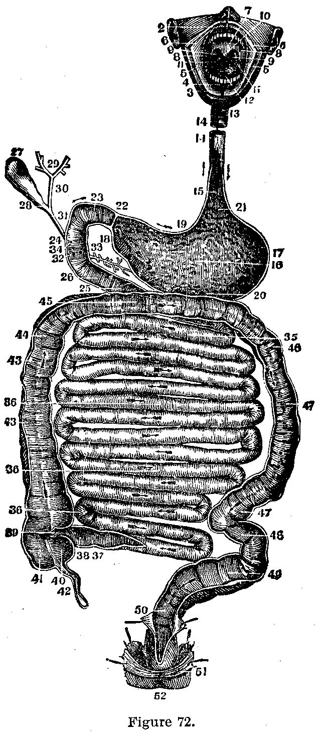
What it Consists of.—The digestive apparatus consists of the alimentary or food canal (Fig. 72) which extends from the mouth to the anus and is between twenty and thirty feet in length, and of the various glands which open into it. The alimentary canal is divided into the mouth, pharynx, esophagus, stomach, small intestine and large intestine. Its function is to digest or convert the food into an assimilable form.
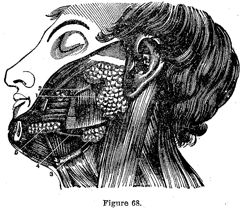
The Mouth.—The mouth is an oval cavity containing the tongue and teeth. In it the food is ground up and mixed with saliva (Fig. 68), which not only moistens it so that it may be readily swallowed but acts on the starchy foods changing them to sugar. The teeth are described in the chapter on teeth. The saliva is secreted by the parotid gland below and in front of the ear, and by the submaxillary and sublingual glands which lie in the floor of the mouth.
|
|
|
|
|
|
|
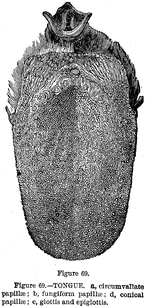
The Tongue.—The tongue (Fig. 69) is a muscle covered by mucous membrane, containing many mucous glands and little projections called papillae in which are lodged the ends of the taste nerves. The tongue is the organ of taste, assists in articulation, and aids in mixing the saliva with food and keeping the food between the teeth.
The Pharynx.—The pharynx is really the upper part of the esophagus, expanded into a muscular bag. It hangs from the skull above, is four and a half inches long, and communicates with the nose, ear, mouth, esophagus and larynx.
The Gullet.—The esophagus or gullet is nine inches long, of same construction as the pharynx, and empties into the stomach. After the food is chewed it is forced into the pharynx which contracts and pushes it down into the esophagus, which propels it onward to the stomach. During swallowing the opening into the larynx is closed by a little trap door called the epiglottis.
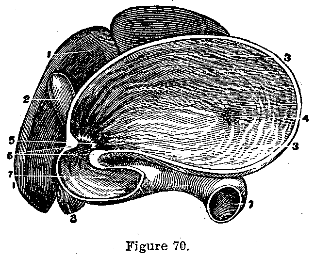
The Stomach.—The stomach (Fig. 70) is roughly pear-shaped; the big end, lying on the left side, measures twelve inches transversely and four inches vertically, is situated just below the diaphragm, receives the esophagus in its upper left wall (cardiac opening), and empties into the small intestine at the extreme right (pylorus), the opening being guarded by a circular valve reinforced by muscular fibres.
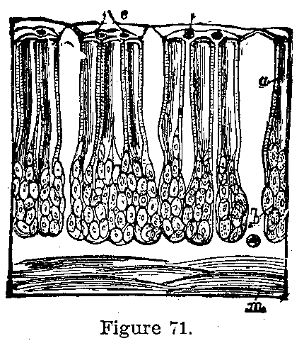
The Stomach Walls.—The stomach wall is made of four layers: externally the peritoneum prevents friction; next is the muscular coat which churns the food, then the cellular coat which carries the blood-vessels, and internally is the mucous membrane (tripe of cow) containing thousands, of little glands (Fig. 71), the peptic or gastric glands, which secrete the gastric juice. When food reaches the stomach, the cardiac orifice and pylorus close, the stomach contracts and mixes it with the gastric juice which is now freely secreted.
|
|
|
|
|
|
|
The Gastric Juice.—Gastric juice is made of water, salts, hydrochloric acid and pepsin. It changes albumen to peptone, which is readily absorbed, dissolves the cellulose of vegetable and the fibrous tissue of meats. Water and some of the peptones are absorbed by the stomach. The remaining portion of the food, now a liquid, passes on into the intestines.
Small Intestine.—The small intestine is about twenty feet in length, one inch in diameter, and extends from the stomach to the cecum, into which it empties. It is connected to the spine by a fold of peritoneum, the mesentery, and is contained in the lower and central portion of the abdomen. It is divided, beginning above, into the duodenum, jejunum and ileum.
Duodenum.—Into the duodenum empty the ducts of the liver and pancreas. The small intestine has four coats similar to those of the stomach. In the small intestine the albumens are changed to peptones, fat emulsified, and starches converted into sugars by the action of the bile from the liver, the pancreatic juice and the intestinal juices. The peptones, fat and sugar are absorbed by the intestinal walls and the remaining portion of the food passes into the large intestine which also absorbs to a slight extent the nutritious portions of its contents, which are now senusated and are called feces.
|
|
|
|
|
|
|
Large Intestine.—The large intestine is five feet in length, runs from an enlarged pouch, the cecum, into which the small intestine empties, to the anus. It is about three times as large in calibre as the small intestine. The cecum is situated in the right lower corner of the abdomen, ending below in the vermiform appendix which varies from three to six inches in length and is about one-quarter inch in diameter. In the cecum the large intestine ascends to the liver (ascending colon), passes over to the spleen on the left side (transverse colon), descends on the left side (descending colon) to the pelvis where it curls like an S (sigmoid flexure) and then ends in the rectum which reaches the surface of the body as the anus. As the feces accumulate in the large intestine they are forced downward to the sigmoid flexure and rectum where they remain until expelled from the body.
|
|
|
|
|
|
|
The Sweetbread.—The pancreas (sweetbread) is a long narrow gland about seven inches in length lying behind the stomach. Its duct opens in common with the bile duct, into the duodenum.
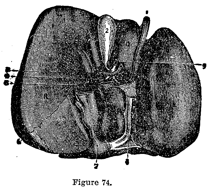
The Liver.—The liver (Fig. 74), the largest gland in the body, weighs about four pounds, and is situated in the upper right corner of the abdomen, where it is retained by the peritoneum which after forming its outer coat, runs to the abdominal walls as ligaments. It is divided into five lobes, which are made up of lobules, each about one-twentieth of an inch in diameter, between which the vessels and ducts ramify. The bile duct has appended to it a pear shaped bag, the gall bladder, which, lying on the under surface of the liver, acts as a reservoir for the bile during the intervals of digestion. The bile duct unites with the pancreatic duct and empties into the duodenum. The liver secretes bile, stores up sugar from the blood, helps make blood, destroys poison in the blood and excretes urea and allied products.
|
|
|
|
|
|
|
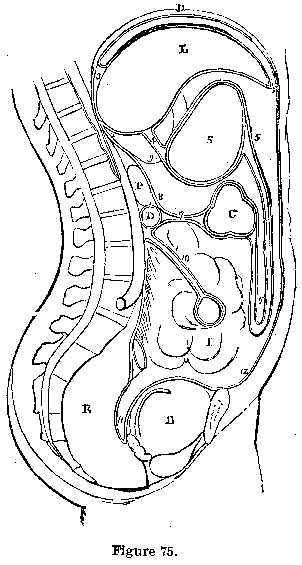
Peritoneum.—The peritoneum (Fig. 75) covers all the abdominal organs; it is a serous sac containing a small quantity of fluid which prevents friction between the organs it covers. The omentum is a double fold of peritoneum, which falls from the front of the stomach nearly to the bladder, then ascends to the transverse colon.
This page is maintained by
Charles Keith.
Contact:
Send me a message
Last Modified: Monday, 13-May-2013 15:31:46 EDT