|
|
|
|
|
|
|
Absorption.—Absorption means the passage of materials from mucous surfaces, serous cavities or tissues into the lymph or blood-vessels.
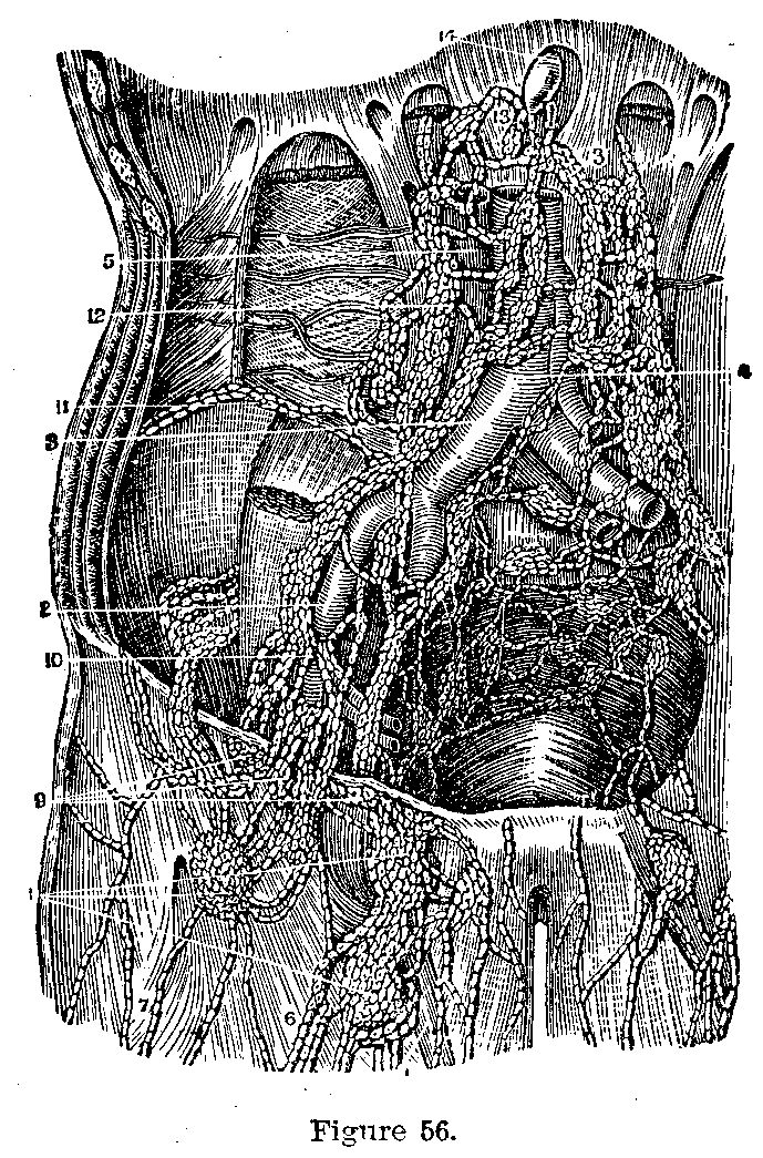
The Lacteals.—The digested fat in the intestines is absorbed by lymph vessels, called lacteals, because their contents resemble milk. These lacteals converge from various parts of the intestine to form the thoracic duct which passes up and empties into a large vein is the neck. The dilated lower end of the thoracic duct is called the receptaculum chyli. The lymph from the tissue all over the body is collected into the lymphatic vessels (Fig. 56) which, finally by two big trunks, the thoracic duct and the right thoracic duct, into the veins of the neck. On its way to the blood the lymph passes through the lymph glands which frequently swell when any poison passes through them, the kernels felt in the neck during an attack of tonsilitis for example.
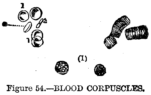
Composition of Blood.—The blood is made from the food we eat, and it in turn feeds all the tissues of the body and drains away all their waste products. It consists of liquor sanguinis (liquid of blood) and corpuscles (little bodies), the former containing water, proteids, salts, nutritive and excrementitious matter. The corpuscles (Fig. 54) are red, which are 1/3200 of an inch in diameter, circular and biconcave, or white, which exist in the proportion of one to three or four hundred reds, are 1/2500 inch in diameter and possess amoeboid motion. When blood is exposed to air it clots, a stringy material proteid in nature, fibrin, which exists in solution in the liquor sanguinis, entangles the corpuscles forming a semisolid mass.
|
|
|
|
|
|
|
Function of Corpuscles.—Blood corpuscles carry oxygen from the lungs to the tissues and the liquor sanguinis carries food; the blood drains carbon dioxide and other waste products from the tissues to the excretory organs: skin, kidney, liver and lungs.
Circulatory Apparatus.—The blood is carried to and from the tissues by the circulatory apparatus which consists of the heart, arteries, capillaries and veins. The heart pumps the blood through the arteries to the thin-walled capillaries where the food passes out to the tissues and waste is given to the blood; from the capillaries the blood drains into the veins which run to the heart. The heart then sends the blood to the lungs to be purified, to the intestines for food and again pumps it out to the tissues.
The Heart.—The heart is a hollow muscular organ of conical form, placed in the chest between the lungs and inclosed in a serous sac, the pericardium. It is placed obliquely; the base, to which is attached the great vessels, is directed upward and backward; the apex is directed downward and to the left, and corresponds to the interval between the fifth and sixth ribs, one inch to the inner side and two inches below the nipple.
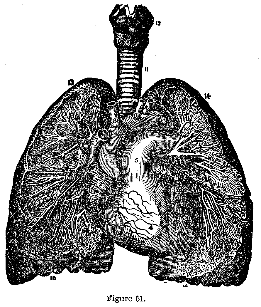
In a grown person (Fig. 51) the heart is about five inches in length, three and a half inches in breadth at its broadest part and two and a half inches thick. In the male it weighs from ten to twelve ounces and in the female about two ounces less.
Heart Divisions.—The heart is divided longitudinally by a muscular partition into two halves and a transverse partition divides these halves into two cavities. The lower cavities are called ventricles and the upper ones auricles. The walls of the auricles are thinner than those of the ventricles and the walls of the right side of the heart are thinner than those of the left.
|
|
|
|
|
|
|
Right Auricle.—The right auricle receives the blood from the two main veins of the body—the two vena cavae. From the auricle the blood is forced into the right ventricle through the auriculo-ventricular orifice. This opening is guarded by the tricuspid valve, to prevent the reflux of blood into the auricle when the ventricle contracts. This valve is composed of three segments, to the free margin of which are attached tendinous cords, which, springing from the muscular ridges projecting from the inner surface of the ventricle, the columnae carneae, give support to the valves.
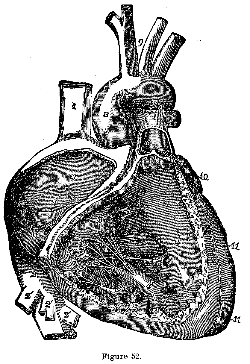
Right Ventricle.—The walls of the right ventricle (Fig. 52) are about one-third as thick as those of the left ventricle. Beside the opening into the auricle there is the opening into the pulmonary artery which is guarded by the semilunar valves, three semicircular folds of the lining membrane of the heart.
Course of the Blood.—The blood is forced from the right ventricle through the pulmonary artery to the lungs, the semilunar valves closing after each contraction of the ventricle so preventing any backward flow.
|
|
|
|
|
|
|
Left Auricle.—The left auricle is smaller than the right, but thicker; it receives the blood which returns from the lungs by the pulmonary veins and forces it into the left ventricle through an opening, guarded by valves, similar to the right auriculo-ventricular orifice, except that the valve, called the mitral valve, has but two segments.
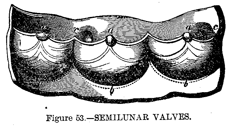
Left Ventricle.—The left ventricle is the thickest and strongest portion of the heart. The blood received by it through the auriculo-ventricular orifice is discharged into the main artery of the body, the aorta, through an opening which is guarded by semilunar valves (Fig. 53) as in the case of the pulmonary artery. The cavities of the heart are lined by a delicate endothelium, which is continuous with that of the blood-vessels.
|
|
|
|
|
|
|
Pulsation.—The heart pulsates from seventy-five to eighty times per minute in the adult; in childhood it is more rapid. The strength and rapidity are governed by the nerves which supply the heart with force.
Heart Sound.—Upon listening to the heart two sounds are heard. The first sound, dull and heavy, is caused by the contraction of the heart, the shutting of the auriculo-ventricular valves and the rush of blood. The second sound, sharp in character, is due to the snapping shut of the semilunar valves.
Function of Arteries.—The arteries carry the blood from the heart to all parts of the body. It has three coats, an outer areolar elastic coat; a middle muscular coat and an inner endothelial coat.
The Aorta.—The main artery of the body is called the aorta (Fig. 43). It springs from the left ventricle, runs up toward the neck, then turns and descends along the spine and divides in the lower abdomen into the two common iliac arteries.
Coronary Arteries.—Just after leaving the heart it sends the two coronary arteries to the heart muscle. Then as it arches, through the chest it gives off. on the right side the innaminate artery and on the left side the left common carotid and the left subclavian arteries. The innominate divides into the right common carotid and subclavian arteries.
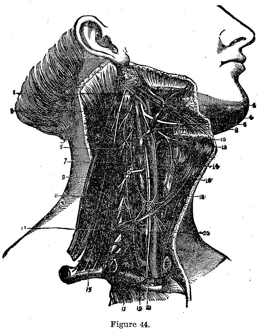
Cardiacs.—The carotids (Fig. 44) run up the neck to the top of the larynx where they divide into the external carotid which supplies the outside of the head and the internal carotid which supplies the brain, ear and eye.

Subclavian.—The subclavian (Fig. 45) supplies the chest, neck and upper extremity; when it reaches the armpit it is called the axillary artery and in the arm it is called the brachial. This trunk, called subclavian axillary and brachial, according to its situation, gives off numerous branches to the various structures of the shoulder and arm. At the elbow it divides into the radial and ulnar branches. The brachial lies on the inner, protected side of the arm just beneath the biceps muscle. It is important to know its location when making pressure to stop hemorrhage lower down the arm.
|
|
|
|
|
|
|
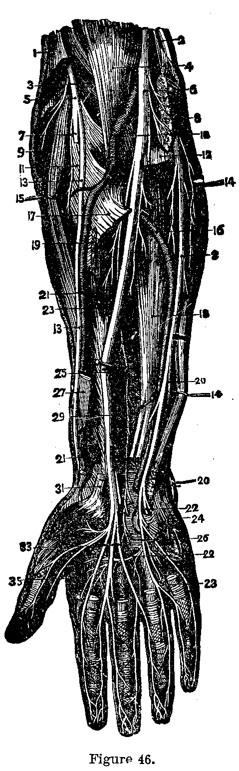
Radial Artery.—The radial artery (Fig. 46) from the bend of the elbow down the radial side of the arm to the wrist, where it is frequently felt to determine the character of the pulse; it then winds around the base of the thumb, enters the palm between, the thumb and metacarpal bone of the index finger and forms an arch (deep palmar arch), which sends branch to the thumb, index finger and palm.

Ulnar Artery.—The ulnar artery, larger than the radial, passes. down the inner side of the forearm, giving off branches to the muscles. In the palm it also describes an arch (superficial palmar arch) which sends branches to the fingers.
Thoracic Aorta.—The portion of the aorta in the thorax is called the thoracic aorta, that in the abdomen the abdominal aorta. The thoracic aorta supplies the pericardium, lungs, esophagus and intercostal structures with nourishment.
|
|
|
|
|
|
|
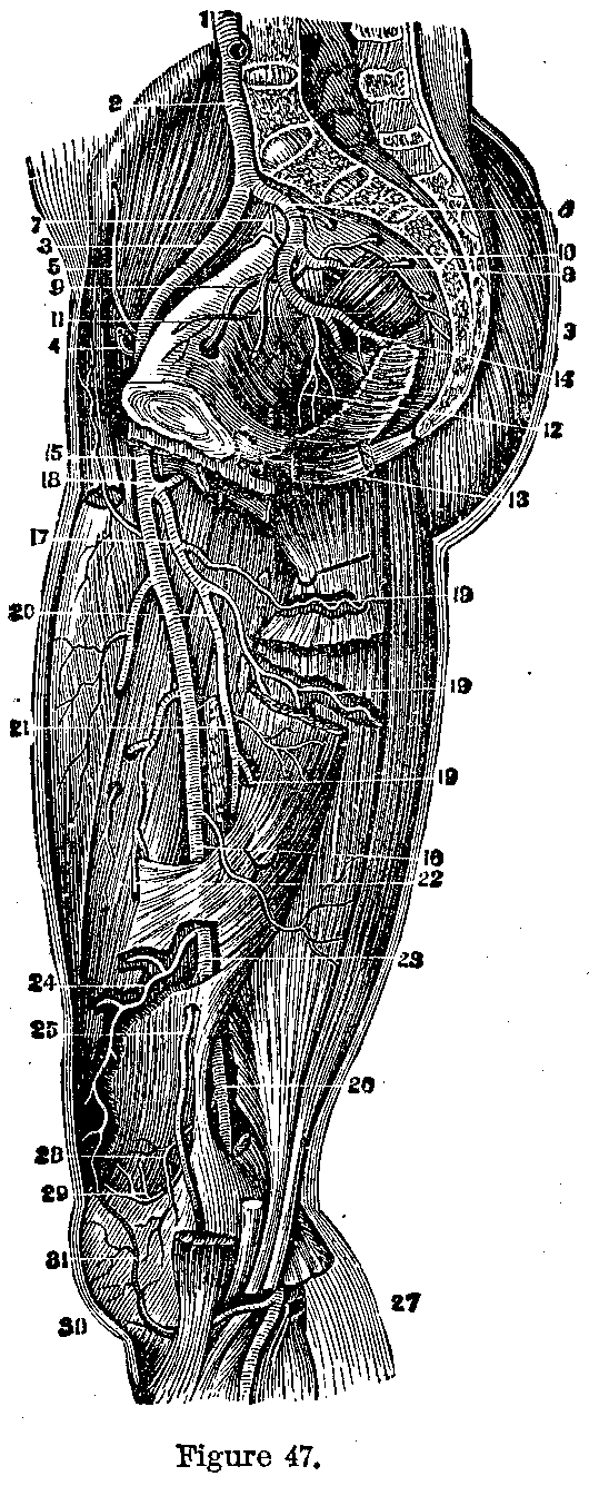
Abdominal Aorta.—The abdominal aorta (Fig. 47) supplies the diaphragm,stomach, liver, spleen, intestines, kidneys, ovary or testicle and muscles of the abdominal wall by branches whose names correspond to the organ it supplies.
Common Iliacs.—Opposite the fourth lumbar vertebrae the aorta divides into the two common iliacs, short trunks which again divide into the internal and external iliac arteries, giving off no branches.
The internal iliac dips into the pelvic cavity and divides into two trunks; the anterior gives off branches to the bladder, rectum, anus, genital organs, buttocks and upper part of the thigh; the posterior trunk sends branches to the buttocks, sacrum and muscles in the pelvis.
|
|
|
|
|
|
|
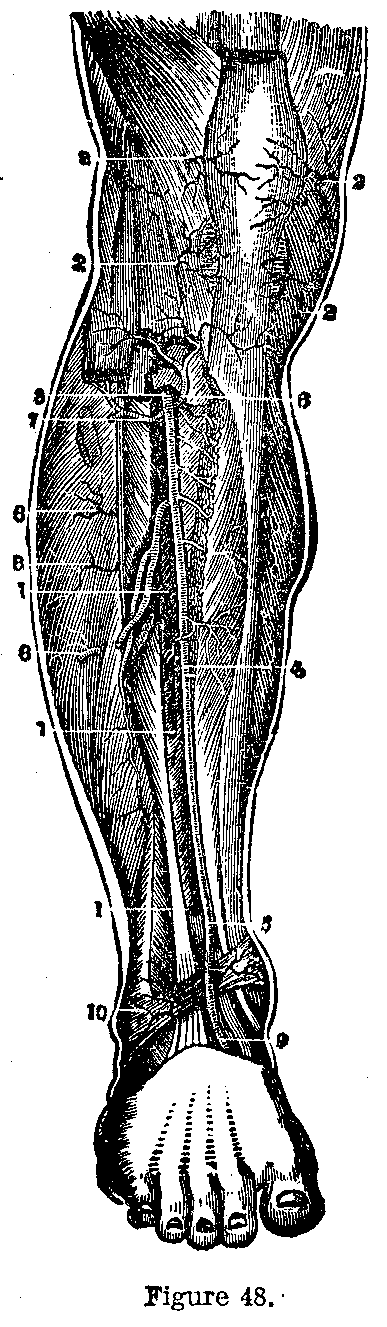
External Iliac.—The external iliac (Fig. 48) runs across the pelvis and escaping below Poupart's ligament is continued down the thigh as the femoral artery. It gives off two large branches to the muscles of the belly.
|
|
|
|
|
|
|
Femoral Artery.—The femoral artery runs a straight course down the thigh from the middle of the groin to the lower third of the femur, where it passes through an opening in the muscles and becomes the popliteal. After giving off several small vessels to the muscles of the thigh it sends out a large trunk, the profunda, which gives off two, the circumflex and three perforating branches, which supply the muscles.
This page is maintained by
Charles Keith.
Contact:
Send me a message
Last Modified: Monday, 13-May-2013 15:31:46 EDT