|
|
|
|
|
|
|
Short Bones.—Short bones are placed where strength is more necessary than mobility, as in the hand and foot; their structure is spongy, covered by a thin layer of compact bone.
|
|
|
|
|
|
|
Flat Bones.—The flat bones are found where protection of important organs is necessary, as in the skull, sternum and scapulae. They consist of two tables of compact bone filled in with cancellous tissue. Certain bones do not belong to one class alone and are called mixed bones.
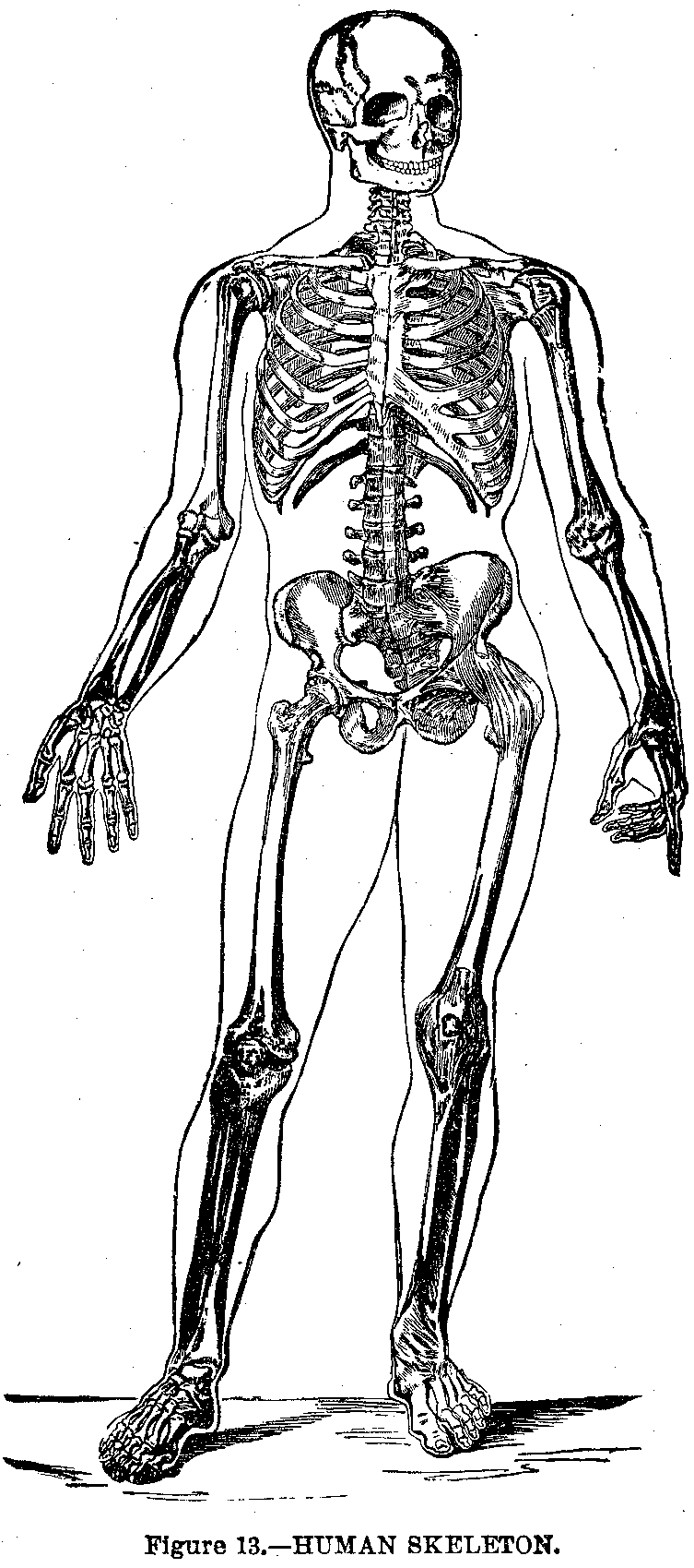
Number of Bones.—There are in the adult skeleton, excluding the teeth, ossicles of the ear, and Wormian bones, 200 separate bones. These are:
In the spinal colum.............. 26
In the skull.....................  8
In the face...................... 14
Ribs, breast bone and hyoid bone. 26
Upper extremity ................. 64
Lower extremity ................. 62

Spinal Column.—The spine (Fig. 14) is a flexible column made of small bones called vertebrae, seven cervical, twelve dorsal, five lumbar, five sacral and four coccygeal vertebrae.
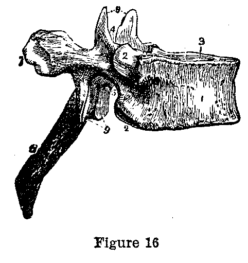
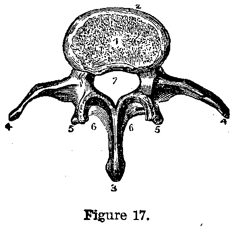
Spinal Vertebrae.—A vertebra (Figs. 16 and 17) consists of a solid portion in front called the body, and an arch behind, so that when placed one above the other as in Figure 14, the bodies of the vertebrae form a support for the body and the arches a canal which contains and protects the spinal cord. The arches are formed by a plate of bone on each side (lamina) joined to the body or constructed portion of bone (pedicle) and unite behind to form the spinous process, which is the portion one feels when running a finger down the back.
|
|
|
|
|
|
|
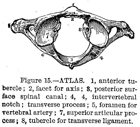
The Atlas.—The first cervical vertebra or atlas (Fig. 15) has neither body nor spinous process, but consists of an anterior and posterior arch and two lateral masses on which rests the skull. The axis, or cervical vertebra has a projection from the upper surface of its body (odontoid process) which fits in the anterior arch of the atlas, permitting the head with the atlas to be rotated from side to side.
The Sacrum.—The sacrum consists of five vertebrae welded into one bone. It is triangular in shape and is wedged in between the haunch bones forming the back of the pelvis. Attached to its apex is the coccyx, which consists of four vertebrae so joined as to form one bone.
Ligaments.—The vertebrae are tied together by ligaments. Between each vertebra and its neighbor is a disk of cartilage which acts as a buffer to prevent shock and allows the spine to bend in various directions.
Bones of the Skull.—The skull is divided into the cranium or brain case and the face. The cranial bones are one occipital, two parietal, one frontal, two temporal, one sphenoid and one ethmoid. The occipital bone forms the back and under part of the skull. It is perforated by a large opening (foramen magnum) which transmits the spinal cord to the spinal canal. The cerebellum rests on its inner or upper surface, the external surface give attachment to muscles.
Side and Top Bones.—The parietal bones form the sides and top of the cranium, joining in the median line and being placed between the occipital bone behind and the frontal bone in front.
Frontal Bone.—The frontal bone forms the forehead and forms the roof of the orbit on the upper surface of which rests the brain. The orbital plates are separated by the ethmoid bone, which is spongy and filled with perforations which transmit the nerves of smell to the nose.
|
|
|
|
|
|
|
Temporal Bone.—The temporal bone consists of a squamous or scale-like portion which overlaps the parietal bone, and a petrous or stony portion which helps form the floor of the cranium. The petrous portion lodges in the internal and middle ear.

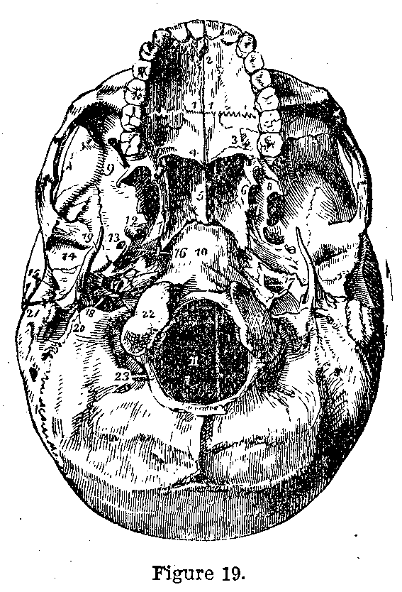
Sphenoid Bone.—The sphenoid resembles a butterfly in shape. It is the keystone of cranial architecture binding the bones of the head firmly together.
Facial Bones.—The facial bones are: two nasal bones, forming the bridge of the nose; two superior maxillary (upper jaw); two lacrymal, forming a portion of the inner wall of the orbit; two malar or cheek bones; two palate bones forming the back part of the roof of the mouth and the corresponding portion of the floor of the nose; two inferior turbinated bones which are scrolls of bone placed in either nostril; one vomer, forming the partition between the nostrils; and the lower jaw or inferior maxillary bone, horseshoe in shape, joining with the temporal bone above and being freely movable below to permit chewing, talking, etc.
|
|
|
|
|
|
|
Hyoid Bone.—The hyoid bone is U-shaped, situated in the neck just above the larynx and gives attachment to many muscles of the tongue and throat.
The Chest.—The thorax or chest is an elastic bony cage made by the breast bone in front, the spine behind, and the ribs and their cartilages at the sides. It is filled by the heart and lungs, which it protects. The sternum or breast bone occupies the middle line anteriorly, is flat, and is made of three pieces, the manubrium ( handle), the gladiolus (blade), and the pointed extremities, the ensiform or xiphoid appendix, these names were given by the ancients who compared it to a sword.

The Ribs.—There are twenty-four ribs (Fig. 20), twelve on each side. They are joined to the vertebra behind and to the sternum, by means of cartilages, in front. They are irregularly semicircular in shape, flattened antero-posteriorly, and slightly twisted on themselves. The head joins the vertebra behind, the neck is the constriction in front of the head and the angle, the point of greatest curvature. The seven upper ribs unite directly with the sternum and are called true ribs. The other five are called false ribs, the upper three being united in front to the cartilages of the ribs above them, and the last two having no attachment in front are termed floating ribs.
The Shoulders.—The upper extremity consists of the. shoulder, the arm, the forearm, and the hand. The bones of the shoulder are the clavicle and scapula connecting the arm with the trunk. The clavicle, collar, or key-bone is a short bone curved like the letter f running horizontally between the sternum and scapula.
|
|
|
|
|
|
|
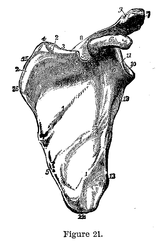
The Scapula.—The scapula (fig. 21) forms the back of the shoulder, is triangular in shape, the apex pointing downward and lies on the ribs. On the upper part of the outer surface is a thick triangular spine, the outer extremity of which (the acromion) forms the point of the shoulder.
Socket of the Shoulder.—Beneath this process the upper angle is hollowed out to relieve the upper end of the arm bone. In front of this depression is a curved prominence, the coracoid process.
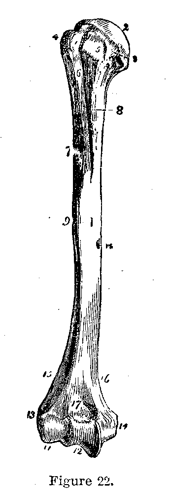
Arm Bone.—The humerus (Fig. 22) or arm bone consists of a long cylindrical shaft, having a rounded head above for articulation with the scapula, and a broad flattened lower extremity for articulation with the forearm bones.
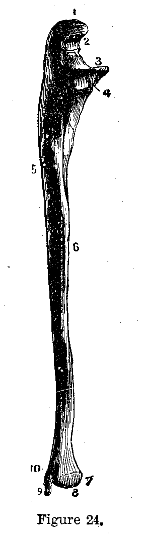
The Forearm.—The bones of the forearm are the ulna and the radius. The ulna (Fig. 24) lies on the inner side of the forearm when the palm of the hand faces upward. The upper extremity which joins the humerus, has two processes, the olecranon, forming the point of the elbow and the coronoid process, which complete the hinge joint of the elbow in front. The lower end of the ulna is small and does not articulate with any bone of the wrist.
|
|
|
|
|
|
|
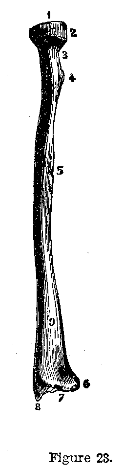
The Radius.—The radius (Fig. 23) lies on the outer side of the forearm. It has a cup-shaped head for articulation with the humerus. The rounded edge of the head fits in a concavity of the ulna and is surrounded by a sling-like ligament, which allows the head to rotate. The lower extremity is larger and is hollowed out to fit the wrist bones.
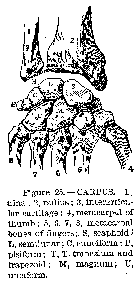
The Wrist.—The wrist or carpus consists of eight small bones in two rows (Fig. 25). In the upper row, beginning at the radial side, are the scaphoid, semilunar, cuneiform, and pisiform, bones; in the lower row, the trapezium, trapezoid, or magnum and unciform bones.
The Hand.—The hand is made of five short cylindrical bones called the metacarpal bones (Fig. 25), to the lower extremity of which are attached the finger bones or phalanges, there being two for the thumb and three for each finger.
This page is maintained by
Charles Keith.
Contact:
Send me a message
Last Modified: Monday, 13-May-2013 15:31:46 EDT