|
|
|
|
|
|
|
Popliteal Artery.—The popliteal is the continuation of the femoral running in the hollow behind the knee joint, dividing just below the knee joint into the anterior and posterior tibial arteries.
Anterior Tibial.—The anterior tibial passes forward between the bones of the leg at its upper part, passes down the front of the leg, and, on the front of the foot, becomes the dorsalis pedis.
Dorsalis Pedis.—The dorsalis pedis runs along the back of the foot and terminates in the artery of the great toe; it gives off branches to the tarsus and metatarsus, the latter forming an arch and giving branches to the toes.
Posterior Tibial.—The posterior tibial descends along the inside of the back of the leg to the hollow behind the inner ankle, where it divides into the two plantar arteries. It gives branches to the muscles of the leg, tibia and ankle.
Plantars.—The internal and external plantar arteries crossing the foot form an arch, from which branches are given to the toes, in a manner analogous to those in the hand.
Pulmonary Artery.—From the right ventricle of the heart arises the pulmonary artery, which conveys the impure blood to the lungs to be purified.
Vein Function.—After the blood flows through the capillaries it is collected by the veins, which are made by small branches joining to form larger branches and these again joining larger trunks, and so on.
Vein Structure.—The veins have three coats, like the arteries, but are thinner, less elastic, and when empty collapse. They have valves at intervals to prevent the backward flow of blood.
Jugular Vein.—The small veins of the exterior of the head follow the arteries and have similar names. They empty into the external jugular which runs down the neck and empties into the subclavian vein.
Internal Jugular.—The internal jugular receives the veins from the interior of the cranium, passes down the neck with the carotid artery and unites with the subclavian vein to form the innominate vein.
Innominate Veins.—The two innominate veins are in the chest and Join to become the superior vena cava.
Veins of Upper Extremity.—The veins of the upper extremity (Fig. 49) besides those accompanying are a radial, an anterior and posterior ulnar and a median vein. They collect the blood from the hand and forearm, and just above the bend of the elbow the ulnar veins unite to form the basilic vein, which passes up the inner side of the arm and empties into the axillary vein.
|
|
|
|
|
|
|
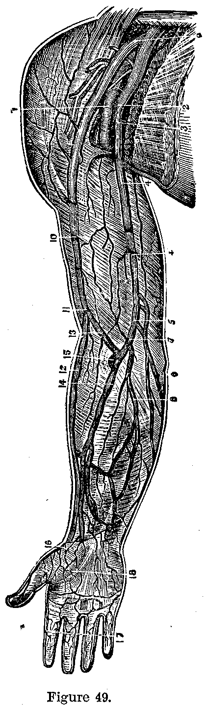
Radial Vein.—The radial vein forms the cephalic, which passes up the outside of the arm and winding around the shoulder-joint empties into the axillary vein.
Median Vein.—Below the bend of the elbow the median and a branch from the deep veins empty into a large Y-shaped vein; running from its apex into which these veins empty it unites together the basilic and cephalic veins, one arm being called the median basilic and the other the median cephalic.
|
|
|
|
|
|
|
Axillary Vein.—The axillary vein, in the armpit, runs up to form the subclavian which joins the internal jugular to form the innominate.
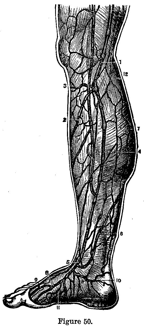
The internal saphenous vein (Fig. 50) commences on the back of the foot, and running straight up the inner side of the leg and thigh joins the femoral vein just below Poupart's ligament.
Saphenous Vein.—The external or short saphenous vein begins at the outer side of the foot, runs up the middle of the calf of the leg and empties into the popliteal vein.
The deep veins follow the arteries and have similar names.
Femoral Vein.—The femoral vein receives all the veinous blood from the leg, runs with the femoral artery into the abdomen, becomes the external iliac which joins with the internal iliac to form the common iliac. The two common iliacs join to form the inferior vena cava which runs up the spine and empties into the right auricle, receiving in its course the various abdominal veins.
Portal Vein.—The veins from the stomach, spleen and intestines are collected into a short trunk, the portal vein, which enters the liver. The blood from the liver is collected by the hepatic vein which empties into the inferior vena cava.
Pulmonary Veins.—The four pulmonary veins start as capillaries in the walls of the air cells of the lungs, carry pure blood and empty into the right auricle.
In order to reach the lungs air passes through the nose, pharynx, larynx and trachea, which warm it and filter it of impurities.
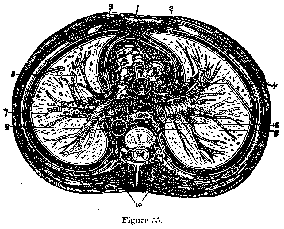
The Larynx.—The larynx (Adam's apple) is the organ (Fig. 55) of voice, and will be described in connection with disease of the throat.
Windpipe.—The trachea (windpipe) is made of rings of cartilage, joined by connective tissue. It is five inches long and lies just beneath the skin of the neck until it enters the chest where it divides into the two bronchial tubes, one going to each lung. These divide and subdivide into numerous branches.
The Lungs.—The lungs, are conical, slate colored in adult life and are separated In the middle of the thorax by the heart, gullet and great blood-vessels. The outer surface of the lungs is convex and smooth, the inner surface concave. Above it extends into the neck, below it rests upon the diaphragm. The right lung is the larger and is divided into three lobes, the left into two.
|
|
|
|
|
|
|
Lung Lobes.—Each lobe is made of little lobules which consist of a little ramification of a bronchial tube communicating with air cells.
Lung Membrane.—The surface of the lung is covered by a smooth serous membrane, the pleura, which is reflected upon the walls of the chest; the intervening space contains a small quantity of fluid which prevents friction during the respiratory movements.
Breathing.—When the chest is enlarged by elevation of the ribs and descent of the diaphragm the lungs follow the chest wall and expand, air rushing into them. When the muscles relax the elastic and over-distended lungs discharge the air through the windpipe.
Air Vesicles.—In the air vesicles the blood is separated from the air by a very thin partition through which oxygen passes to the blood corpuscles. The expired air contains the carbon dioxide and other impurities with which it has been charged while in the air vesicles.
Oxyginized Blood.—The blood, after passing through the lungs, is a brighter red, richer in oxygen, cooler and is rid of its impurities.
Function of Nerves.—The nervous system presides over all functions and harmonizes them. It permits the environs to be recognizable. It may be compared to a telegraph system, of which the brain is the central station, to a rider on a horse, or to the captain of a steamship. It is divided into the cerebro-spinal system (brain and spinal cord with their nerves) which presides over the animal functions, motion, sensation, etc., and the sympathetic system which controls the organic functions, nutrition, growth, etc. The sympathetic system is composed of a series of ganglia ((large mass nerve cells) in the head and along the front of the spine, connected by nervous cords.
|
|
|
|
|
|
|

The Brain.—The brain is a huge mass of white, and gray nervous matter contained in and protected by the cranium. It is surrounded by three membranes (meninges): the dura mater, externally, dipping into the fissures to form the falx cerebri, tentorium cerebelli and falx cerebelli which separate and support portions of the brain; the arachnoid, the serous membrane, supplying a fluid which acts as a water cushion for the brain; and the pia mater, the layer carrying the blood-vessels.
|
|
|
|
|
|
|
Weight of Brain.—The average weight of the brain is fifty ounces in males, and six ounces less in females.
Divisions of Brain.—The brain is divided into the cerebrum, cerebellum, pons varolii and medulla oblongata.
The Cerebrum.—The cerebrum is the largest part of the brain, resting on the roof of the orbit, base of skull, and tentorium cerebelli. It is divided into lateral halves by the falx cerebri. The halves are joined by the corpus callosum. Internally it is composed of white, and externally of gray, nervous tissue. The gray tissue is wrinkled into convolution and is the active portion of the brain, the white matter conducting the nerve impulses to and from it. It is the seat of memory, intelligence, reason, will, motion and sensation.
|
|
|
|
|
|
|
The Cerebellum.—The cerebellum lies beneath the posterior portion of the cerebrum, is gray outside and white inside. It coordinates muscular movements.
The pons varolii connects the various parts of the brain. It conducts impulses to and from the brain.
Medulla Oblongata.—The medulla oblongata is the enlarged upper end of the spinal cord resting in the cranium. It is made of blended white and gray tissue, conducts the nerves from the brain to the spinal cord and contains independent nervous centres which regulate the heart, lungs, blood-vessels, sweating, etc.
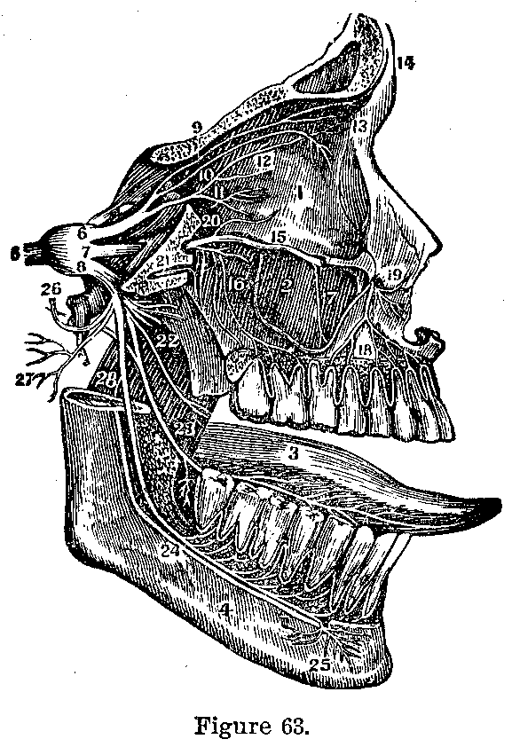
Nerves of Brain.—The brain gives off twelve nerves on each side. The nerves pass out through holes (foramen) in the skull and supply the organs of sight, smell, taste and hearing, and also motion and sensation to certain parts. They are: the olfactory (smell), optic (sight), motor oculi (motion to eye), patheticus (motion to superior oblique muscle of eye), trifacial (sensation to face, motion to chewing muscles and nerve of taste), abduces (rolls eye out), facial (motion to face), auditory (hearing), glossopharyngeal (taste and sensation), pneumogastric (presides over swallowing, heart, lungs, etc.), spinal accessory (motion to muscles, neck and back) and the hypoglossal (motion to tongue).
|
|
|
|
|
|
|
Spinal Cord.—The spinal cord is a long tail hanging from the back of the brain and contained in the spinal canal. It is sixteen to eighteen inches long and surrounded by three membranes like the brain.
|
|
|
|
|
|
|
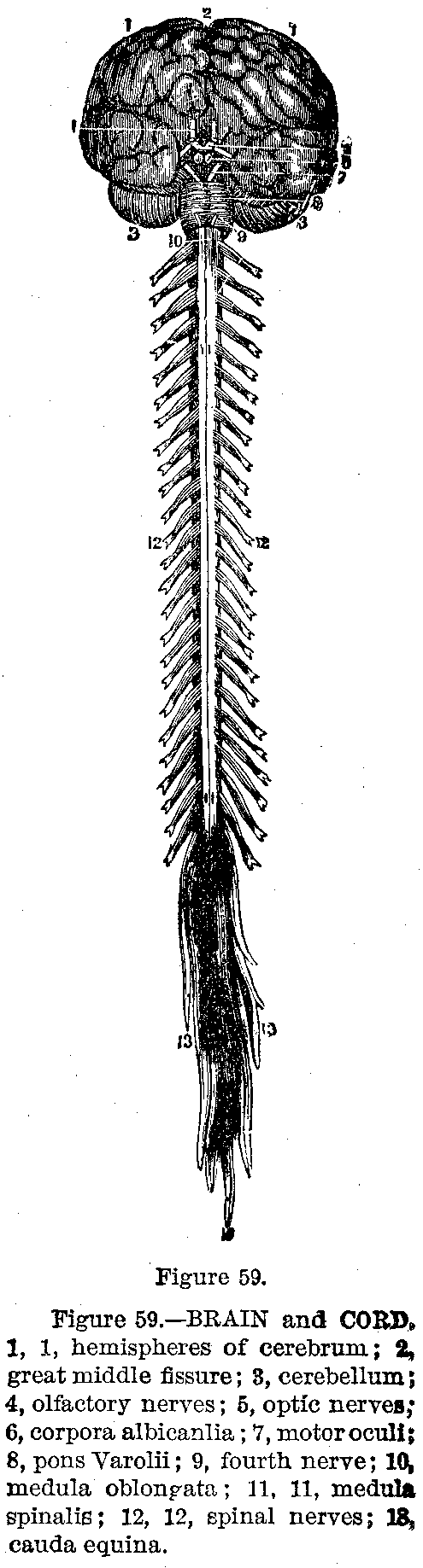
Upper Section.—Upon section the cord (Fig. 59.) is seen to be composed externally of white nervous tissue, and internally of the gray, which is arranged somewhat in the shape of the letter H. The cord is divided by two antero-posterior fissures into two equal lateral halves, which are united in the centre by a bridge of gray matter.
The spinal cord is a great nerve cable carrying fibres to and from the brain; it. also coordinates motion, presides over the nutrition of certain parts and contains independent nervous centre.
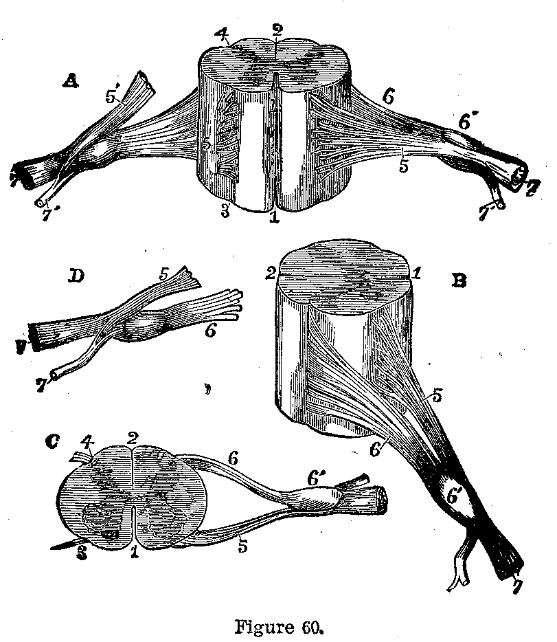
From the anterior horn of the gray matter arises the motor roots of the spinal nerves, and from the posterior horn the sensory roots.
Spinal Nerves.—Each spinal nerve, of which there are thirty-one pairs, consists of the anterior or motor and the posterior or sensory root. These unite within the spinal canal and form a single cord (Fig. 65), which passes through the opening between the vertebrae and divides into two trunks, one for the anterior and the other for the posterior surface of the body.
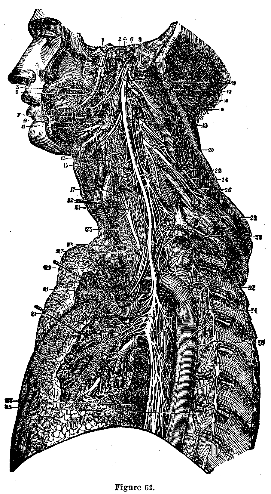
Cervical Plexus.—The anterior branches of the four upper cervical nerves unite with. each other to form the cervical plexus which gives on branches to the side of the head, neck, shoulder, chest and diaphragm. The anterior branches of the fifth, sixth and seventh cervical nerves unite, the fifth receiving a branch from the fourth; the eighth cervical and first dorsal nerves unite; these cords form the brachial plexus, and after sending nerve trunks to the muscles of the neck and sides of the chest below the collar bone, these two trunks each send off a trunk which unite to form a third or posterior trunk which divides into two branches, supplying the muscles and skin of the outside and back of the arm, forearm and hand.
|
|
|
|
|
|
|
Inner and Outer Trunks.—The inner and outer trunks are continued down the inside of the arm, and again each sends a branch to form a middle cord, the median nerve. The external cord then becomes the musculo-cutaneous, and the internal, the ulnar.
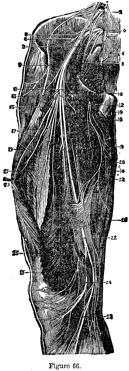
Median Nerve.—The median nerve (Fig. 66) gives off branches to' the muscles and to the skin of the hand. The ulnar nerve is placed on the inner side of the arm and supplies the forearm and hand. The musculo-cutaneous supplies the skin and muscles of the forearm and wrist.
Spinal Nerves.—The twelve dorsal spinal nerves send branches along the ribs and supply the muscles of the back.
Lumbar Nerves.—The five lumbar nerves send posterior branches to the muscles of the back; the anterior branches unite to form a plexus which sends branches to the muscles of the belly and the genital organs; the largest branch, the crural nerve, is distributed to the front of the thigh.

|
|
|
|
|
|
|
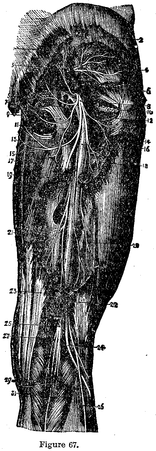
Sacral Nerves.—The fifth lumbar joins the sacral nerves to form the sacral plexus; its largest branch is the great sciatic which passed down the back of the thigh dividing at the knee into the external and internal popliteal nerves; these are continued down the leg as the anterior and posterior tibial nerves supplying the leg and foot.
|
|
|
|
|
|
|

This page is maintained by
Charles Keith.
Contact:
Send me a message
Last Modified: Monday, 13-May-2013 15:31:46 EDT