|
|
|
|
|
|
|
Bones.—The skeleton is the framework of the body, and is composed of an articulated assemblage of hard organs, the bones.
It serves to preserve the shape of the body; forms cases for the protection of the vital organs, and gives attachment to muscles and forms levers of movement.
Number of Bones.—The number of distinct pieces or bones composing the skeleton varies at different periods of life. Some remain distinct from the first moment of their development, such generally being of the simplest form, such as the bones of the carpus or wrist, and the patella or knee cap. Others, which are viewed as single bones in the adult, not only consist of several pieces in the beginning, but in the progress of development have other pieces successively added, as in the case of the vertebrae or bones of the spine and the thigh bones.
May Unite Bones.—Again bones considered as distinct pieces when the body has arrived at maturity, at a later period may become united with those which are contiguous, as in the coössification of the cranial bones. Therefore in the adult skeleton the number of bones to which we usually refer are two hundred and six, exclusive of the teeth, and sesamoid and wormian bones, which are not uniform in number. Of this number twenty-six are found in the backbone or spine; twenty-two in the skull and face; the ribs count twenty-four, twelve on each side, whether the person be man or woman; each arm has thirty-two bones, and each leg has thirty. These bones vary very much in size, shape and thickness, and all have been named and described with great minuteness by anatomists.
Composition of Bones.—The bones under every modification of shape and mechanical arrangement are constituted by precisely the same elementary matters, the principles of which are an animal and an earthy substance, in intimate combination.
|
|
|
|
|
|
|
Phosphate of lime is the most abundant mineral material, being about 51 parts in the 100 of bone. Carbonate of lime, 11.3 parts; fluoride of calcium, 2 parts.
The animal matter of bone is gelatinous, allied to cartilage; originally every bone is developed from cartilage by ossification.
The mineral matter of bone increases with age; making the bones of the old more brittle. There is more of it also in some bones and parts of bones than in others.
Structure of Bone.—A good idea of the structure of a bone may be gained by picking the second joint of a chicken or turkey clean, and then sawing off about an inch of the upper end and splitting this piece in half lengthwise with a hatchet or strong knife. This thigh-bone of a turkey corresponds to the femur or thigh-bone in a human being, where it lies near the centre of the leg and reaches from the hip to the knee. Its upper end may be recognized by its having upon it a single round knob, which formed part of the hip-joint.
Marrow of the Bone.—On examining such a bone it is found to be hollow in the middle, and partly filled with a fatty substance called the marrow of bone. In this marrow run important little blood-vessels, which carry the blood to nourish the bone, and from it they pass and repass to minute channels running lengthwise in its substance, which are called the Haversian canals, after the name of the physician who first discovered them. In dried bones, and especially when they have undergone prolonged bleaching in the open air, such as those of a long dead horse or cow, these Haversian canals may be readily seen, looking like fine pores in the broken ends of the bones.
Telescopic View of the Canals.—Under the microscope they are discovered to be encircled with rings of lacuna, or little holes in the bone substance, each of which communicates with its neighbors by very minute branching tubes. During the life of an animal the lacuna are filled with soft, jelly-like bone-corpuscles, but in dried bones these gelatinous bodies shrivel up, leaving the lacuna empty, except of air, which, by refracting light differently from what the solid bone does, makes the lacuna look like black dots under the microscope.
Bones of a Chicken.—If the breast-bone of a young chicken is examined it will be found that its smaller end is made up of the tough, milk-white or semi-transparent substance called gristle or cartilage. When a chicken is first hatched nearly all its bones are chiefly composed of cartilage, and as it grows older they gradually become more and more ossified, that is, changed into bone. But certain portions of the body commonly
(Continued on page pg0613)
|
|
|
|
|
|
|
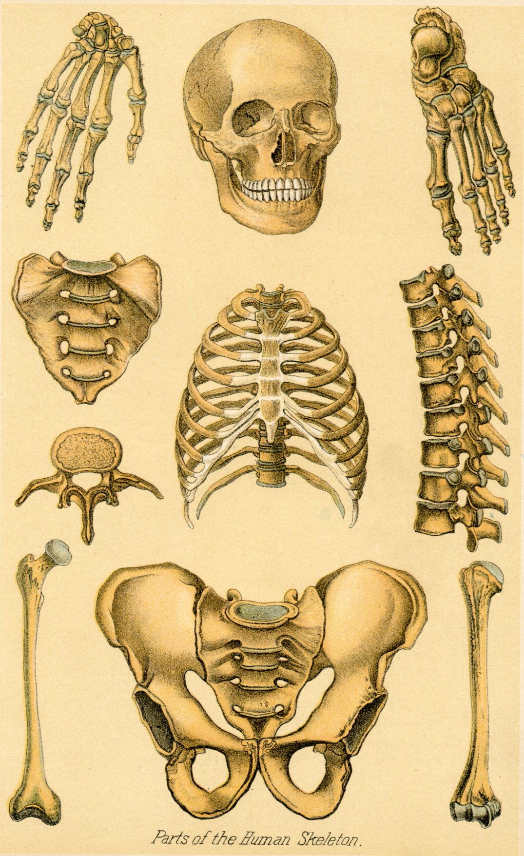
Top Central Plate.—This shows the bones of the cranium, or head, eight in number. The large frontal bone forms the forehead. The articulation of the teeth are prominent. It also shows the facial bones, or those of the face, fourteen in number.
Middle Central Plate.—This shows the bones of the chest; the sternum, or breast bone, in the centre; the ten true and two floating ribs on either side, and part of the backbone, to which the ribs are attached.
Lower Central Plate.—This plate shows the bones of the pelvis. Pelvis means basin. It is the basin or girdle by which the bones of the lower body, as the hip bones, are joined to the upper. The two large side bones are the os innominata, or unnamed bones. The central triangular bone is the sacrum, a composite bone, forming the union between the vertebrae and os coccyx, or tail end of the backbone.
Upper Left-Hand Plate.—This represents the eight bones of the carpus (wrist); the five of the metacarpus (between wrist and phalanges), and fourteen bones of the phalanges (battle rank bones), twenty-seven in all.
Next Figure Below.—This is the sacrum, detached from the pelvic girdle. It is also seen in lower middle plate. It is called sacrum (sacred), because it was of old offered up in sacrifices.
Third left-Hand Figure.—This is a section, or single joint of the backbone, or spinal cord.
Bottom left-Hand Plate.—This represents the femur, or great thigh bone. It is cylinder-shaped, and the largest, longest and strongest bone in the body.
Upper Right-Hand Plate.—This represents the bones of the foot. They are divided into three classes. 1. Tarsus, or ankle bones, seven in all. 2. Meta-tarsus (between tarsus and phalanges), five in all. 3. Phalanges (battle-rank bones), fourteen in all, a total of twenty-six foot bones.
Middle Right-Hand Plate.—This represents a part of the backbone or spinal column. It consists of a series of vertebrae, within which is the spinal cord or nerve, to injure which means paralysis or death.
Lower Right-Hand Plate.—This is the humerus (shoulder), or bone of the upper arm. It is the only bone in the upper arm, and it revolves on the scapula (shoulder-blade) above, and the ulna and radius of the forearm below.
|
|
|
|
|
|
|
remain cartilaginous through life, as, for instance, the grisly bands which fasten the ribs to the breast-bone, and the rings of the trachea or windpipe.
Bones of Arm.—The bones of the arm, counting from the shoulder downward as it hangs at the side, are first the humerus or arm-bone, which extends to the elbow, and next the two bones of the fore-arm, which make up the part from the elbow to the wrist, and are named the radius and ulna. The latter of these two bones projects on the under side of the arm, and the radius has the hand attached to it, and is rolled part way around the ulna every time the hand is turned over from the position of pronation, or lying with its palm downward, to that of supination or lying upon its back. This is a very important movement, and great care must be taken by the use of properly padded splints to save it, when the radius and ulna of the fore-arm are fractured by accident, as very frequently happens.
Bones of Wrist.—The wrist or carpus is composed of eight small bones, each with a hard name derived from Greek or Latin, and the palm of the hand is formed by four of the metacarpal, a word meaning beyond the wrist bones, the metacarpal bone of the thumb making the fifth. The bones of the fingers consist of three rows of phalanges, the thumb having two phalanges only.
Beside the bones mentioned, anatomists reckon the shoulder-blade, or scapula, and the collar-bone, or clavicle, as belonging to the arm, or, as they name it, the upper extremity.
Kinds of Joints.—The joints of the arm exemplify the two chief kinds of articulations made use of in the human body, namely the ball and socket joint, of which the shoulder is an example, and the hinge-like joint, of which the elbow is a good illustration. The joints between the metacarpal bones and the first row of the phalanges of the fingers, that is, those at the roots of the fingers, are imperfectly formed ball and socket joints, and allow, as anyone can see in his own hands, of a good deal of lateral or sidewise, as well as backward and forward motion. The other articulations of the fingers are hinge-joints, and like the hinges of a gate, permit only of a back and forth motion.
The Synovial Fluid.—The ends of bones where they rub against each other inside the joints are covered with firm smooth cartilage, and to diminish the friction as much as possible these polished surfaces of cartilage are kept slippery by a peculiar liquid named the synovial fluid or "joint-water." This synovial fluid, called also the synovia, acts the part of oil to a door-hinge, and when sometimes in old people the synovia becomes scanty, their joints will creak and grow stiff, just as the hinges of a door do for want of oil. The ends of the bones in a joint are held in place by a tough, firm wrapper, called the capsular ligament, which encloses them tightly on all sides, and also prevents the synovia from escaping and being lost.
|
|
|
|
|
|
|
Bones of leg.—In general arrangement the bones of the legs are very similar to those of the arms, making allowance for the difference in function of the two pairs of limbs. The thigh-bone or femur is the longest and strongest bone in the body, as might be expected from the larger share of work in walking, running and leaping it has to perform. It is articulated (or jointed) to the pelvis at the lower corner of the body by the hip-joint, a ball and socket articulation, which allows of considerable movement in every direction.
Knee to Ankle.—From the knee to the ankle, the leg, like the fore-arm, is furnished with two bones. One of these, called the tibia, is the shin-bone, forming the front of the leg and the inner side of the ankle, that is, the side next the other limb. The bone on the outer side of each leg and each ankle is named the fibula, and is much smaller than the tibia, its partner in the business of supporting the weight of the body. The ankle or tarsus is composed of seven bones instead of eight, as are found in the carpus, and it is articulated below and in front, near the middle of the foot, with the five metatarsal bones. At the front, ends of the metatarsal bones are jointed on the toes, each with its three rows of phalanges, except the great toe, which, like the thumb, has but two.
Nature of Sprains.—The ankle-joint is more apt to be "sprained" or "strained," than any other, and this accident, therefore, requires a few words of explanation here. A strain of a joint is the result of moving the bones which compose it too far, or in an unnatural direction, so that the capsular and other ligaments are stretched or perhaps torn a little by the force applied. A strained joint is very painful, apt to swell rapidly, and often proves troublesome for months, or even years, if not properly treated. Until a doctor comes, the injured articulation should be placed in an elevated position, so that the blood will drain away from it, kept perfectly quiet, and covered with cloths wrung out of hot or cold water, so as to reduce the danger of inflammation.
Bones of Skull.—The bones of the skull or cranium are broad, comparatively thin, and curved in such a way as to make a hollow case or oval box, shaped a good deal like an egg, for the protection of the brain, which is placed inside of them. The most important are the frontal or forehead bone, the two temporal or temple bones and the occipital bone, which is at the back of the head. These bones are united together by seams or sutures, consisting of a curious kind of dove-tailing, which fastens them so firmly together that, in their natural state, it is impossible to get them apart without breaking them.
|
|
|
|
|
|
|
Character of Skull Formation.—The arched form of the skull makes it much more capable of resisting blows upon the head; but if these are so severe as to fracture the bones, especially if they are dented in so as to press upon the brain, unconsciousness is often very quickly produced. This would happen much more frequently were it not for the layer of spongy matter interposed between the hard plates which form the cranial bones.
Bones of the Face.—The bones of the face are comparatively light and thin, except the lower jawbone, into which are set the lower teeth, and which is the only bone about the head which is furnished with a movable joint, except the occipital bone, where it rests upon the neck.
Bone of Spine.—The spine or backbone is made up of twenty-four verebrae, the sacrum and the coccyx. These latter bones seem to be each composed of several vertebrae, which, for the purpose of being rendered stronger, have grown fast together. The uppermost vertebra is called the atlas, because upon it the head is supported; and the second is named the axis, because upon it the atlas, and with it the whole head, turns, as in shaking the head negatively. The upper seven vertebrae are called the cervical or neck bones; the next-twelve are designated as the dorsal or back vertebrae; and the last five are named the lumber vertebrae or vertebrae of the loins. These twenty-four bones are fitted together in such a way as to form a continuous tube, which receives and protects the spinal cord or spinal marrow just as the upper continuation of the spinal cord— that is, the brain—is encased and protected by the bones of the skull. The vertebrae are jointed so as to allow considerable motion, both sideways, forward and backward, and have between each pair a cushion of fibrocartilage, which serves to preserve the brain from injury by the shocks and jars which would otherwise be given to it in jumping, running and various other movements. The sacrum, which is continuous with the vertebrae, is united with two large, flat and irregularly-shaped bones, to form the pelvis or basin at the lower part of the trunk. The pelvis supports the spine and the organs in the abdominal cavity, and is in its turn sustained on each side by the thigh bones, which prop it up at the hip-joints, as already indicated.
Bones of the Thorax or Chest.—The heart and lungs are protected by a bony cage composed of the twenty-four ribs, which lie a little beneath the skin of the thorax or chest and in thin persons can be easily felt at the sides or near the breast-bone. This breast-bone or sternum is situated directly in the middle or front of the thorax and has the front ends of the ribs attached to it by cartilages, named the costal or rib-cartilages, which allow of the outward and upward movement of the ribs, necessary in breathing.
|
|
|
|
|
|
|
The back or posterior ends of the ribs are jointed on to the vertebrae of the spinal column in such a way as to allow of needful motion, and yet secure sufficient stability and firmness.
Dislocation of Bones.—When a dislocation occurs, or, as it is commonly called, a bone is put out of joint, the bones composing an articulation have been pulled or twisted so hard as to displace them, breaking some of the ligaments which are arranged to keep them in their proper positions.
Example of Dislocation.—Generally, a person whose arm, at the shoulder, for example, is dislocated, suffers a good deal of pain and loses the use of the limb until the bones are put back in their places again, or as it is called, the dislocation is reduced.
A dislocation is one of the heavy penalties people often have to pay for imprudent over-exertion in lifting and wrestling, or for carelessly letting themselves have falls and hard knocks, or becoming entangled in railway accidents.
Repair of Fractures.—The repair of broken or fractured bones is a wonderful process of nature, in which a material called callus, at first like putty, is formed around the broken ends, holding them together, feebly at first, but afterward it gradually hardens, uniting them more firmly indeed.
Since this "knitting" of the broken bones may occur in almost any position they happen to lie in, or are pulled into by the irritated muscles in the neighborhood, it is evidently very important that they should be put and kept in exactly the right place. For this purpose there are many ingenious splints and bandages devised and used by surgeons, as is pointed out in chapter on Wounds and Accidents.
Inflammatory Affections of the Bones and of their enveloping membrane (the periosteum) are occasionally met with, sometimes following injuries and wounds of bone (and microbic infection), but are much more common as a result of syphilitic or scrofulous disease than as a primary complaint.
|
|
|
|
|
|
|
Symptoms.—Pain is an early and persistent symptom usually worse at night; there is gradual swelling of the affected part, tenderness on pressure, and the skin over the inflamed area becomes red and edematous. If the process goes on in the diseased bone the centre of the portion involved may die or undergo necrosis, as it is termed, just as local death may occur to connective tissue; for example, the slough or core of a common boil.
Nodes.—These are hard bony swellings, the result of a subacute inflammation not going on to caries or necrosis, which are apt to occur as tertiary symptoms of constitutional syphilis.
They are most common in long bones and are frequently found on the front surface of the tibia or shin-bone. They are often very painful for a time, particularly at night, but yield promptly to treatment with the Iodide of potassium in the majority of cases.
Softening of Bone (Osteomalacia).—This is an uncommon disease observed in adults; seldom seen in males, occurring in greater frequency in females. It is characterized by a softening of the bones, rendering them very liable to break or bend on the application of little force; resulting from absorption or deficiency of the earthy matters in the bones. An analogous condition is sometimes found in the insane, and has no doubt given rise unjustly to some of the stories of cruel treatment in asylums for these unfortunates.
Brittleness of Bone (Fragilitis Ossicum).—This is an affection of bone in which the inorganic are out of proportion to the organic constituents, rendering the bones brittle; there is an apparent increase of the earthy matters, with a diminution of the vascularity of the bone. Children and young persons seem to suffer most from this disease, and in many instances an hereditary tendency can be traced. It is probably due to defective innervation.
Osteoma (Chondroma).—The bones are subject to tumors, to cartilaginous or other growths. They form hard rounded tumors, fixed to their point of origin. They may attain a huge size—but are usually small. They grow slowly without pain or other symptoms except such as may be caused by their bulk or pressure, and interfere with the functions of neighboring nerves.
Osteomata are liable to inflammation and necrosis, but never undergoes malignant or cancerous degeneration. Chondromata may not only become inflamed, and necrosis and sloughing follow; but it becomes cancerous in some instances; for instance, after an injury to a bone, a chondroma may appear and develop with terrible rapidity, and upon its removal may return, become cancerous and form secondary tumors elsewhere.
|
|
|
|
|
|
|
Treatment.—If osteomata or chondromata are removed whilst small the operation is to be recommended, but when left until a huge tumor has developed, of the nature of which no doubt can be entertained, any interference is of questionable propriety.
Synovitis.—Among the important diseases of the joints must be mentioned synovitis or inflammation of the lining membrane, by which the synovia or joint-water is secreted. This disease, usually attended with severe pain, and when the joint is a large one accompanied with much constitutional disturbance and fever, appears in two forms, the acute and the chronic.
Causes.—The acute form is usually the result of injury, which may be very slight in its character, as even the least puncture of the joint by which the air can enter is liable to produce it. Acute synovitis runs its course in ten or twelve days, causing much swelling and severe suffering on the slightest movement of the limb to which the joint is attached. The chronic form is commonly a continuation of the acute, and may itself result in softening and what is called pulpy degeneration of the synovial membrane.
Treatment.—The treatment of synovitis is by free leeching of the affected part; perfect rest in bed, with the limb elevated and secured in a splint if needful; low diet with saline purgatives, such as epsom salts or seidlitz powders, and anodynes to relieve pain. In the chronic form small blisters and painting with tincture of iodine are likely to prove useful. Inflammation of the synovial structure of the joints is apt to take on a rheumatic, scrofulous or syphilitic character in persons who are constitutionally under the influence of these taints. In such instances the appropriate treatment for them, as already pointed out, is to be associated with that for ordinary synovitis.
Ulceration of the cartilages may occur in a joint as a consequence of long continued inflammation, causing intense pain, and usually disabling the limb. Its liability to occur renders the prompt treatment of synovitis doubly important.
Abscess (Coxalgia).—Abscess within a joint is rare in healthy persons, but in the scrofulous it is by no means uncommon, and in strumous children abscess in the hip-joint, causing the lamentably frequent affection, coxalgia, is a malady of much importance.
Treatment.—The general treatment in these sad cases is that already indicated for scrofula, but the local trouble should be immediately attended to by an experienced surgeon, and remedied as far as possible by the aid of the complicated apparatus devised for the purpose.
|
|
|
|
|
|
|
Pott's Disease.—This consists of a tubercular inflammation of the bodies of the vertebrae or spinal bones, and their cartilages; and is most common in children between two and ten years of age, although it may occur at any age. In some cases the affection appears to follow a slight injury to the spine in those of tubercular or strumous tendencies, in others the disease develops without apparent exciting causes.
Symptoms.—Rigidity of spine, tenderness and local pain are the prominent early symptoms. Abscess may occur early, but is most common in the late stages. Deformity or spinal curvature usually occurs as a result of the disease process, depending upon the amount of breaking down in the bones and the falling together of the vertebrae, and may be gradual or rapid in its development. Treatment is as indicated in article on Coxalgia.
Muscular Function.—The power which moves different parts of the frame, according to the directions of the will, as, for instance, the legs, in walking, is produced by the contraction of muscles. These muscles form the lean meat of animals and of the human body, and, except in very fat people, make up a larger portion of the bulk of the frame than any of its other constituents.
Composition.—They are composed, as is readily seen in a piece of fresh beef or mutton, of long strings of reddish material, which, under the microscope, are found to be made up of a multitude of fine, beaded threads, arranged in small bundles, and called the ultimate muscular fibres. They exercise power in moving the limbs, and so forth, by shortening up or contracting when excited by the nervous fluid, sent to them through the nerves from the brain, as ordered to do so by the will.
Mechanism.—The exact mechanism of moving the arm, for instance, by the process of contraction, may be easily understood from the picture shown on page 621, in which it is readily perceived that the shortening up of the muscle must pull up the hand, bending the arm at the elbow-joint, and changing the position from that represented in the second, to that shown in the third figure. Precisely the same kind of operations accomplish the motions of lifting the feet in walking or climbing, swinging the arm in throwing a ball, opening and shutting the mouth, and, indeed, of most of the voluntary movements of which we are capable.
|
|
|
|
|
|
|
Voluntary and Involuntary.—But whilst many of the muscles are controlled by the will and are, therefore, called voluntary, many of them an not so ruled, and hence have received the name of involuntary.
Most Important Involuntary Muscles.—Among the most important of the involuntary muscles are the heart, the intercostal muscles, the muscles between the ribs which help to expand the lungs in respiration, and the muscular fibres of the alimentary canal, which aid in pushing along the food in digestion. Fortunate is it for us that such is the fact; for otherwise, when our wills were off duty, as in sound sleep, the operations of these vital organs would stop, and life, which depends upon them, soon cease.
Muscular Attachment.—The muscles are usually attached to the bones, and move them by sinews or tendons, which are made of white, fibrous tissue, the strongest and most flexible material in the body. These tendons are like long, round, white cords, such as may be seen in the lower part of the leg of a chicken. The largest tendon in the human body is that of the heel, called the tendon-Achilles, which is the continuation of the big muscle of the calf of the leg. This powerful muscle is used in jumping and, since it acts at a great disadvantage, is necessarily very strong in order to be able to throw the entire body forward, as in making a leap.
Origin and Insertion.—As a general, but by no means universal, rule, a muscle has one attachment which is fixed, commonly spoken of as its origin, and a second, called its insertion, upon which it acts by drawing it toward the origin when the muscular substance contracts. Muscles mostly pass in a straight line between their two attachments, but sometimes they act around an angle by sliding over a pulley, or by means of a small bone in the tendon, like the knee-pan. The muscles are so attached that they are always slightly on the stretch, and thus, at the moment they begin to contract, they are in an advantageous position to bring their action to bear on the bones which they move. When the contraction ceases, the bones are drawn back to their former position without any sudden jerk.
Muscular leverage.—The muscles commonly act upon the bones as levers, by working upon the short arm of the lever, so that more direct force is required on the part of the muscle than there is weight in the body moved. From this arrangement, however, the indispensable advantage is gained that the small contraction of the muscle causes an extensive movement of the part acted upon, and much greater rapidity of motion is secured. Each of the three orders of levers is met with in the different bones of the human skeleton; often, indeed, all three varieties are found in the same joint, as, for example, the elbow, where the simple flexion and extension of the biceps and triceps muscles, which are large, fleshy masses on the front and back of the arm, between the shoulder and elbow, afford excellent illustrations, as shown in the accompanying figure.
|
|
|
|
|
|
|
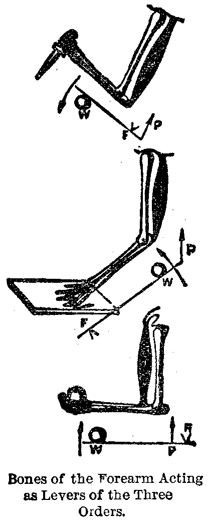
Illustration of Leverage.—The arm is used as a lever of the first order when the triceps is caused to contract, and by pulling upon the upper end of the small bone of the forearm moves the hand around the elbow-joint, which serves as a fulcrum. This is shown in the upper diagram in which the hand is represented as striking a blow with a dagger.
Again the arm furnishes an example of a lever of the second order, when the hand resting on a point of support, such as a table, acts as the fulcrum, and the triceps muscle pulling on the upper end of the ulna or smaller arm bone, by straightening the arm lifts a weight placed upon it in front of the elbow.
The third order of levers is exemplified by the arm when bent by the contraction of the biceps in ordinary flexion of the elbow. Here the muscle, which is the power, is placed between the fulcrum, which is the lower end of the humerus at the elbow-joint, and the weight, which is lifted in the palm of the hand, as shown in the lower diagram.
Number of Muscles.—The whole number of muscles in the human being is not far from five hundred, mostly arranged in pairs on opposite sides of the head, body or limbs. It is, therefore, manifestly impossible to describe them all in detail here, and yet there are a few which are important enough to require notice.
Important Muscles of the Face.—Among the muscles of the face should be mentioned those of the eye, six in number, four of which turn the eyeball up or down, inward toward the nose or outward toward the temple, as becomes necessary to see an object distinctly. The muscles of expression are especially attached about the mouth, and produce their effects by puckering up the lips, as in whistling; drawing up the corners and widening the mouth, as in laughing; pulling down its angles, as in weeping, and so forth. The masseter muscles placed inside the cheeks between the upper and lower jaws are very strong, and enable human beings to chew up some very hard articles of food in the operation of mastication, as has been already explained.
|
|
|
|
|
|
|
Flexor Muscles.—The action of the large muscle of the front of the arm, called the biceps or two-headed muscle, has been already described, The muscles of the fore-arm, which shut the fingers as in clasping the hand, are called the flexors, because they flex or bend the fingers. The tendons by which their power is conveyed may be readily felt on the Inside of the wrist of a man who tries to shut his hand when the fingers are forcibly held open by another person. The fore-arm muscles which open out the fingers after the hand is closed, in doubling up the fist for instance by the flexors, are called the extensors. The tendons of the extensor muscles, when the latter are strongly contracted, show very distinctly on the back of the hand, as straight, hard cords, running from. the root of each finger to the middle of the wrist.
Muscles of the Spinal Column.—The spinal column is almost surrounded, except in front, by a thick mass of muscles, which gives the great strength required by many kinds of laborers, such as porters who carry heavy loads upon their backs. The muscles of the leg, which are needed in walking, running and jumping, are very large and strong, corresponding to the hard work they are called upon to perform.
The Longest Muscle.—The longest muscle in the body is the sartorious or tailor's muscle, which is so named because it helps to bend the lower limbs into the cross-legged posture so frequently adopted by tailors. It lies on the inside of the thigh, is thin and narrow, but sometimes measures over two feet in length.
Intercostal Muscles.—The intercostals are flat, thin layers of muscular fibres, which extend from the lower edge of each rib, except the last pair, to the upper margin of the rib next below. In this way they fill up all the spaces between the bars of the bony cage in which the lungs are contained, and, when they contract, pull up these bars or ribs so as to widen and deepen the cavity of the thorax, and so cause air to be drawn into the lungs.
Inflammatory Disease.—Inflammatory disease of the muscular system, except as it is connected with rheumatism or pyemia, is extremely uncommon. When it occurs it is liable to go on to suppuration, and the formation of abscess. The pain is dull and aching rather than acute, and the disease is to be treated as already directed in speaking of the management of boils.
|
|
|
|
|
|
|
Hypertrophy and Atrophy.—These occur in muscles and produce effects important or otherwise, according to the position of the muscle in the animal economy. The most important hypertrophy is that of the muscle composing the heart, the influence of which has been detailed in the article on valvular disease affecting this organ.
Contractions.—Palsy and spasm in various forms have also been considered in the chapter on diseases of the nervous system, upon which these disturbances chiefly, though not entirely, depend.
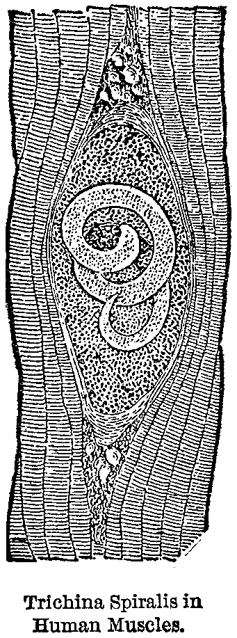
Trichiniasis.—The most important disease of the muscular system not yet discussed is its infection with parasites, and particularly with the trichina spiralis or pork worm, which, in consequence of the late embargoes upon American hams and bacon, has assumed a national or, indeed, an international importance. The great fatality in many cases of trichiniasis, as the malady produced by the trichina has been named, and the ease with which the whole trouble, can be avoided by a proper understanding of the nature and origin of the affection, render a full account of the entire malady and its means of prevention singularly appropriate to a popular work like the present one.
The Trichina Spiralis.—The trichina spiralis, which is next, perhaps, to the echinococcus, the most dangerous animal parasite infecting man, is found also in pigs, foxes, guinea-pigs, rats, cats, mice, marmots, polecats, badgers, and more rarely in some other animals, including the dog. A physician of Philadelphia found that of ten cats which he dissected in 1879 nine were infested with trichinae.
Sources of Trichinae.—Pigs, from which the human race is most apt to become diseased with trichina, are supposed to become themselves infected, chiefly from eating rats, the offal of other pigs, and the excreta of human beings containing trichinae. It is doubtful whether a single case of trichiniasis in man ever occurred where the patients became infected otherwise than by eating raw or underdone pork, and the most common sources of infection are sausages, ham and bacon.
How Trichinae Breed.—If the trichinae existing in diseased pork are very young they may be simply digested, when they reach the human stomach, without being developed. But if the parasites have attained their fuller growth, the cysts which contain them are alone dissolved by the gastric juice, and the embryo is set free. These embryos, after they pass through the pylorus and duodenum, soon become mature, and their thread-like appearance renders them quite easily recognizable by the naked eye. Countless eggs are now discharged by the females, and in about a week's time the new brood of trichinae hatched out from these eggs begin to make their way to the muscles, either by boring their way through the soft tissues, or by being carried along by the current of blood in the blood-vessels, or perhaps by both of these methods of progression through the body. These larval trichinae attain their full size in about two weeks from the time they leave the egg. The males and females are each about one-thirtieth of an inch long and about one seven-hundredths of an inch broad.
|
|
|
|
|
|
|
Effects of Trichinae.—In some few favorable cases severe gastro-intestinal inflammation is set up, and the parasites are violently expelled by diarrhoea, without being able to enter the muscles at all, so that if it were possible to detect the malady with certainty at this stage, nature thus suggests the appropriate treatment by drastic purgatives. In the majority of instances, however, the migration of immense numbers of larval trichinae from the intestinal canal takes place, and occupies in general about four days only. In this brief space of time even the most distant muscles of the body may all be invaded.
Symptoms.—Among the earlier symptoms of the trichina disease are a general feeling of debility and discomfort, and a loss of appetite, to which succeed nausea and vomiting, diarrhoea, prostration of strength, and a sensation of stiffness about the neck, arms and legs. These evidences of illness resemble, it will be observed, to a great extent, the first stage of typhoid fever, for which the cases are usually mistaken if there is no point in the history of the patient to suggest trichiniasis. The further progress of the parasites through the tissues sets up high fever, with frequent pulse and copious offensive perspirations, although the temperature of the body seldom or never reaches the elevation which characterizes that of typhoid. For some days the stiffness of the limbs seems to increase, while all the muscles become painful, swollen, and very sensitive to the touch.
|
|
|
|
|
|
|
A Characteristic Sign.—About the end of the first week the attention of the attending physician is usually awakened (if it has not previously been aroused) to a suspicion of the trichina disease by the appearance of an edematous swelling of the eyelids and root of the nose. This is often the first characteristic sign of trichiniasis, and should be looked for at this period of the illness in all cases of supposed typhoid and rheumatic fever. During the second week movement of the intercostal muscles in respiration grows very painful, thus preventing to a great extent the necessary repose of the patient. If the diaphragm is invaded, severe hiccough is apt to come on, and when the larval trichinae commence to infest the laryngeal muscles hoarseness and loss of voice make their appearance.
Cause Paralysis and Exhaustion.—When a large quantity of trichinous meat has been eaten, so that the immigration into the muscles of the afflicted patient is by millions, they soon cause an almost paralyzed condition, attended by excessive exhaustion. The facial edema continues through the second week, when it generally disappears, and is followed by swelling of the feet and legs, and ultimately of the trunk. At the end of the third week, if the patient survives to this period, the pulse and respiration are very frequent, the tongue is red and dry; the mouth can scarcely be opened, the sweating is profuse, the pain so severe that little or no sleep can be obtained, and there is great anxiety or delirium, death frequently occurring in the fourth or fifth week; with symptoms of profound exhaustion.
Complications.—Complications, such as pneumonia, peritonitis and pleurisy, are not uncommon, but in favorable cases when the number of trichinae is comparatively small, or the constitution of the patient unusually vigorous, the pain, swelling and diarrhoea begin to abate, the oppression of breathing passes off, the desire for food returns, sleep is obtained, and the anemic patient enters upon a slow and tedious convalescence; the parasites having become encysted within the muscles, these gradually become acclimated, as it were, to the presence of the foreign bodies, and slowly regain most or all of their original powers and functions.
|
|
|
|
|
|
|

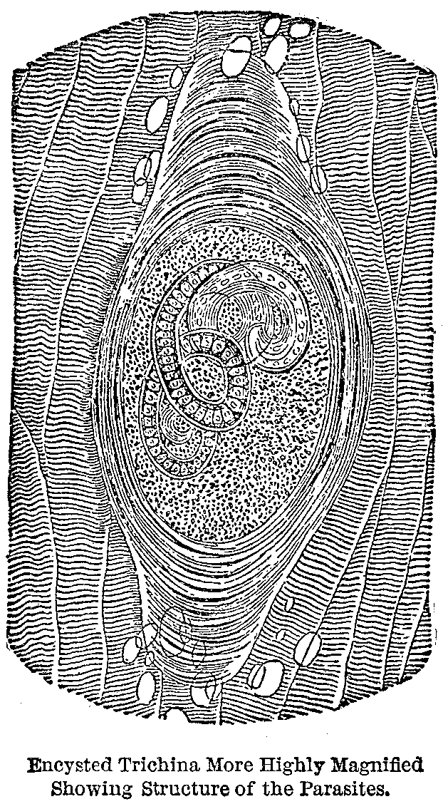
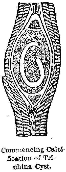
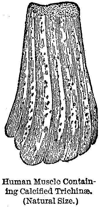
Further Symptoms.—After piercing the fibrous sheath of the muscular fibre bundles, the embryonic trichinae become encysted within lemon shaped capsules (generally one worm in each capsule), of a sort of connective tissue, in which they have some freedom of movement. After a time, these capsules, which, of course, are fixed to one spot in the muscle, become calcified, a process which occupies in man about two years. During this period there is often, for a time, in patients who recover, some loss of power, for a while almost complete, in muscles or groups of muscles; but not infrequently, after this stage is reached, entire recovery ultimately seems to take place. Still, this infested condition of the muscle, which probably exists in thousands of people who walk about utterly unconscious of it, may be the yet undetermined cause of rheumatism and paralysis, or promote the development of consumption and other wasting diseases.
Size of Mature Trichinae Spiralis.—The mature trichinae—the males being about one-eighteenth and the females about one-eighth of an inch long—live in the intestines for six or eight weeks. They never reach the soft tissues of the body, in which their young larvae are so abundant, but are discharged, from time to time, with the excreta of the patient, either alive or after their death has occurred.
Discovery of Trichinae.—Trichinae, as well as cysticerci, were first discovered in human beings in the dissecting room, by Dr. Simon, in 1835. Professor Owen first described and named the trichinae, and Professor Leidy was the first to detect them in the pig. They were, however, repeatedly observed without their true import being ascertained until 1860, when Dr. Zenker, of Dresden, explained their origin and relation to certain symptoms of obscure attacks of sickness, and described the disease trichinosis or trichiniasis. In the same year Leukart published his elaborate and trustworthy investigations upon the subject of the trichina. Previous to 1860 the trichina had been identified only once in pork, although, as occurring in man, it had been well known for a quarter of a century. It is not decided how it will retain its vitality when encysted in human muscles.
Remarkable Vitality of the Trichinae.—Professor Langenbeck, of Berlin, has reported a case where, in removing a tumor from the neck of a patient, eighteen years after the man had an attack of trichiniasis, which passed for poisoning at the time, he found living trichinae in the fragments of attached muscles; and it is stated on good authority that they have been known to exhibit signs of life after a still greater lapse of time. As in this instance, before Zenker's discovery, very many cases passed for poisoning, for typhoid and rheumatic fever, and for other diseases. An epidemic involving over five hundred persons in Blankenberg, Germany, was treated as an outbreak of gastro-rheumatic fever, and it was only several years afterward that the attention of one of the gentlemen who suffered from the disease being called to Zenker's discovery, he submitted to an operation for the removal of a small piece of one of his pectoral muscles, in which encysted trichinae were detected, and the true nature of the disorder which had affected the five hundred patients many years previously was revealed for the first time.
|
|
|
|
|
|
|
Epidemics of Trichiniasis.—In this country severe epidemics have occurred in New York, Mississippi and Iowa, and isolated cases are from time to time appearing in various other States. In 1875, it is stated, that there were some eighty cases of trichiniasis in Berlin, and about seventy-five near Hanover. A group of cases occurred in 1882 in Bridesburg, and another near New York. In 1874 there was quite a severe epidemic in the family of a pork packer residing in Buffalo, New York. The disease is as rare in France as it is common throughout the German Empire.
Vitality of Trichinae After Death of Infected Hog.—In pork the trichinae may be found either encysted or naked among the muscular fibres. It is not certain how long they may live after the pig is killed, but they are known to be capable of propagation after remaining for one hundred days in putrid pork. The frequency of the disease in swine is probably as great, if not greater, in America than elsewhere; but here mankind is not so often infected, because less raw or under-done sausage, ham, bacon, and so forth, is eaten with us than in Europe. Of 1,394 hogs taken at random, and examined by the Chicago Academy of Sciences, twenty-eight were found to be infected with trichinae; but this large proportion can only be accounted for on the supposition that an epidemic among swine was then raging.
Method of Detecting Trichinae.—The editor of the American Journal of Microscopy recommends that in examining the flesh of swine suspected of being infected with trichinae, the following method should be adopted: The parts of the animal to be first tested are the diaphragm, the tenderloin, and the muscles of the head and throat. In the ham, the most likely place to find the parasites is where the muscle ends in tendon. A thin slice should be cut off with a sharp scalpel, or with a pair of scissors curved on the flat. This thin section should then be soaked for some minutes in
(Continued on page pg0629)
|
|
|
|
|
|
|
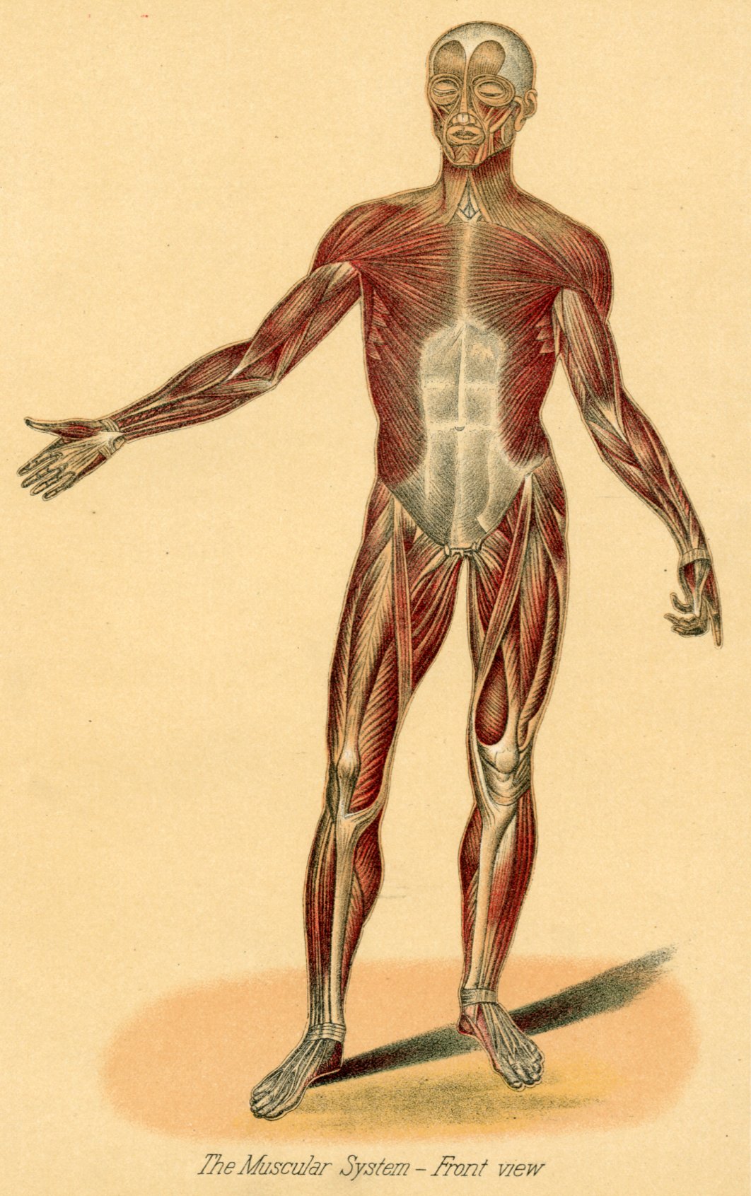
The plate presents a comprehensive view of the front muscles of the body. They may be grouped and viewed thus:
Head Muscles.—1. These are seen above the eyes, and are used for elevating the upper eyelids and corrugating the forehead.
Face Muscles.—2. These are muscles of expression and mastication. Those about the eyes are used in winking and opening and shutting. Those seen at either side of the nose lift the cheeks and lips. Those at the sides control the lower jaw in eating.
Neck Muscles.—3. These serve to lower and raise the head and turn it from side to side.
Shoulder Muscles.—4. These embrace the shoulders and upper arm. They are the great lifting and hitting muscles. The prominent one on the upper arm is the biceps muscle, or muscle with two heads.
Muscles of Forearm.—5. These control rotary, flexor and extensor motion, from the elbow to the wrist.
Hand Muscles.—6. These control all hand motions—opening and shutting, rotation, flexor and extensor movements.
Chest Muscles.—7. These are radiating from sides to centre. They control the twisting, elevation and lowering of the upper part of the body.
Abdominal Muscle.—8. This is seen in the centre, in white. It is intimately connected with breathing and raising and lowering the diaphragm. At its top is the solar plexus, the spot upon which prize-fighters seek to deliver their knock-out blows.
Hip, Thigh and Leg Muscles.—9. These powerful muscles cooperate for every kind of movement and exhibition of strength. The two strap-like muscles of the upper leg are the sartorial, or tailor's, muscles, which enable us to cross our legs.
Lower Leg Muscles.—10. These are also powerful, and possessed of rotary, flexor and extensor power. They largely control the feet in walking, operating clear to the ankle joint.
Foot Muscle.—11. These control from instep to toes, each toe having its elevating and depressing muscle. The rotary motion of the foot is imparted wholly from the ankle.
|
|
|
|
|
|
|
acetic acid, spread out on a glass slide, and covered with a thin glass in the ordinary way; or, if the section happens to be very thick, a compressorium, in which the two plates of glass are forced together by means of a lever and screw, will be found very useful.
A little instrument constructed on the plan of the compressorium, and called a "trichinoscope," is sold to supply the popular demand for home protection against trichinae.
Infected Pork Should be Destroyed.—All pork which has been found to contain trichinae should be seized, condemned and destroyed, either by fire or by strong mineral acids, such as the sulphuric or nitric. Mere burying of poisonous meat of this kind is obviously not sufficient. Moreover, the question as to whether owners of such carcasses should not be compensated for all property confiscated, is well worthy of consideration; because if such was the rule, butchers and dealers would have no inducement for concealment and fraudulent sale.
Only Safe Rule.—In spite, however, of any apparently perfect system of inspection, such as that adopted in Germany, dangerous meat, from some cause or other, will necessarily escape observation, so that the only safe rule for us to adopt, and to urge upon everybody else, is never to eat any pork which has not been thoroughly cooked.
It is not safe to trust to pickling and smoking, even when these processes are combined, as is ordinarily the case, and it must be borne in mind that meat is seldom thoroughly cooked when cut in slices more than an inch thick.
The fact that two dangerous and often fatal maladies like trichiniasis and cysticercus disease, both of which are generally beyond the reach of medical treatment when once they have infected the human system, are not infrequent from eating pork which has been imperfectly cooked, should render the employment of this culinary precaution in regard to all meats universal, especially as it is a safeguard so easily applied.
Myositis.—This affection consists in an inflammation of the voluntary muscles, and many arise from injury to or overuse of a muscle, from gout or rheumatism, from secondary syphilis, or from infection followed by suppuration.
|
|
|
|
|
|
|
Treatment.—The treatment for injury or overuse of muscle is absolute rest of muscle and the local application of anodyne lotions.
If due to rheumatism or syphilis, prompt relief will follow treatment appropriate to these affections.
Degeneration of Muscles.—Fatty degeneration is occasionally observed in muscular tissue—in which the tissue is converted into a fatty granular mass.
Treatment.—By use of passive motion, massage and electricity to improve nutrition of the muscles.
Ossification.—Ossification of a portion of a muscle, or more frequently of its point of insertion into a bone, is occasionally observed as a result of long-continued irritation. Its course is slow and unaffected by treatment.
Tumors.—Muscles may be the seat of cancerous, syphilitic, vascular, cartilaginous or osseus growths. The treatment depends entirely upon their nature; non-malignant growths can often be removed by dissection, cancerous or malignant growths involving muscles of the extremities should be cut out or, as many cases call for, prompt amputation of the limb.
This page is maintained by
Charles Keith.
Contact:
Send me a message
Last Modified: Monday, 13-May-2013 15:31:46 EDT